Illuminating the Hidden World of Exosomes

In 2024, the Nobel Prize in Physiology or Medicine was awarded to Victor Ambros and Gary Ruvkun for their pioneering discovery of microRNAs (miRNAs) and their critical role in post-transcriptional gene regulation. Their work fundamentally reshaped our understanding of genetic control mechanisms by demonstrating how miRNAs act as powerful regulators of gene expression within cells.
Building on this breakthrough, researchers later discovered that miRNAs are not confined within cells but can be transported between them via extracellular vesicles (EVs), tiny, lipid-bound particles like exosomes that range from 30 to 150 nanometers in size. This insight is redefining how scientists understand intercellular communication, with implications for processes such as development, immune modulation, and disease progression.
Far from being passive cargo carriers, EVs have emerged as central players in areas ranging from cancer metastasis to skin aging. However, they remain difficult to study. Traditional optical microscopes lack the resolution to visualize EVs effectively, and conventional labeling methods can disrupt the delicate biological environments being examined. The complexity and variability of EV interactions further complicate real-time observation.
That’s where new technology steps in. Instruments like ONI’s Nanoimager are transforming how researchers study the nanoscale world. By offering unmatched resolution with minimal disruption to native structures, the Nanoimager enables scientists to visualize and decode the previously invisible language of cellular communication across health, disease, and everything in between.
The ONI Nanoimager: A Window into the Nanoworld
The ONI Nanoimager integrates super-resolution imaging, real-time particle tracking, and advanced fluorescence analysis into a compact, user-friendly system. It allows researchers to directly observe EV membrane proteins such as CD63, CD81, and EGFR with spatial resolution as fine as 20 nanometers. Combined with 3D single-particle tracking, the Nanoimager can capture fleeting fusion events between EVs and cell membranes at video-rate speeds.
One of its most powerful features is label-free, in situ analysis. By incorporating methods such as surface plasmon resonance (SPR) imaging and fluorescence lifetime imaging microscopy (FLIM), researchers can analyze EV concentration, activity, and subpopulations without relying heavily on dyes or chemical markers. For example, FLIM can distinguish tumor-derived EVs from those of healthy cells, a crucial step toward targeted therapeutic development.
Further strengthening its versatility, the Nanoimager supports advanced multimodal techniques like direct Stochastic Optical Reconstruction Microscopy (dSTORM) and Photoactivated Localization Microscopy (PALM). These tools allow scientists to study EV membrane curvature and protein distribution at the nanoscale. Additionally, single-molecule FRET (smFRET) enables the exploration of miRNA-protein interactions, offering deeper insights into how genetic information is transferred at the molecular level.
Together, these capabilities make the ONI Nanoimager an essential tool for unlocking the secrets of nanoscale biology.
| Dimension | Technical Specification | Application Value |
| Sensitivity | Single-molecule detection sensitivity down to 100 pM | Detect EV surface markers (e.g., CD63/CD81) |
| Speed | Ultra-high-speed imaging at 1000 frames/second | Real-time capture of EV dynamic fusion processes |
| Compatibility | Supports fixed cells / live cells / tissue sections | Full workflow study of EV secretion–transport–uptake mechanisms |
| Extensibility | Open Python API interface | Custom algorithm development and specialized analysis |
Transforming Exosome Research Across Applications
The Nanoimager is a critical platform for advancing EV research across various scientific fields. With ONI’s EV Profiler Kit, researchers can perform high-resolution single-particle phenotyping and map key biomarkers such as CD9, CD63, and CD81 with nanometer resolution.
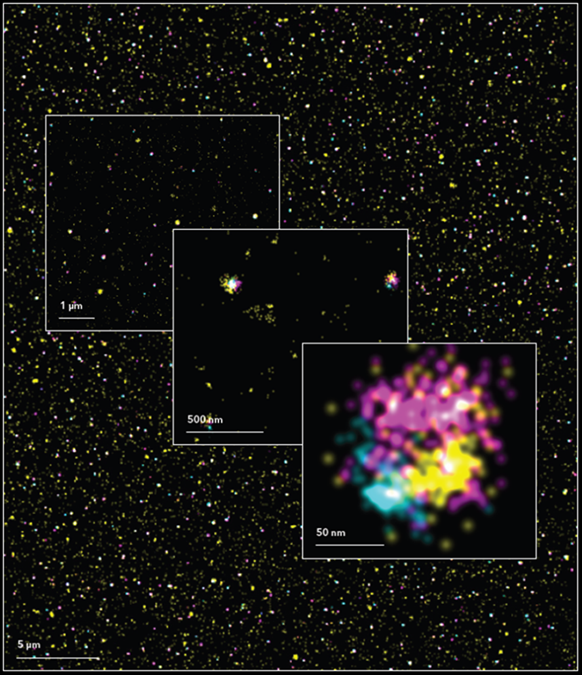
HCT116-derived EV sample captured on ONI’s functionalized chips and labeled with WGA(yellow), anti-CD63-568(cyan) and anti-CD81-647(magenta). Scale bars range from 5μm to 50 nm.
In cancer biology, the Nanoimager captures dynamic interactions between EVs and surface receptors such as EGFR and PD-L1 using time-lapse imaging and single-particle tracking. Techniques like smFRET enable scientists to study how EVs deliver miRNAs to target cells and influence receptor signaling, shedding light on tumor progression.
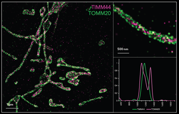
dSTORM image showing inner mitochondrial membrane TIMM44(magenta) and outer membrane TOMM20(green) proteins in U2OS cells, labeled with CF583R and AF647, respectively. Inserts show two examples of individual mitochondria. Line profiles from each mitochondrion were obtained to assess protein distances
In virology and neuroscience, the Nanoimager supports visualization of complex disease models. During the COVID-19 pandemic, researchers collaborated with virologist Stephan Becker’s team to track the movement of SARS-CoV-2 particles and EVs, providing insights into viral spread. In neurodegenerative disease studies, the Nanoimager helps monitor the EV-mediated transfer of toxic aggregates like tau and α-synuclein, advancing our understanding of disease propagation at the nanoscale.
The Nanoimager’s ability to deliver high-resolution, multiparametric data at the single-particle level is propelling breakthroughs in EV biology, disease modeling, and targeted therapeutics.
Beyond the Microscope: A Full Ecosystem for Scientific Discovery
ONI offers more than just powerful imaging; it provides a complete research platform. The ONI Discovery Kit delivers validated end-to-end workflows, and hands-on training ensures fast onboarding and reproducibility.
With the NimOS software suite, researchers can perform cloud-based analysis and collaborate in real time. High-resolution datasets can be securely shared across teams, accelerating experimental timelines and promoting transparency.
The Nanoimager is already powering discoveries across multiple domains:
- Oncology: Optimizing exosome-based drug delivery through detailed EV characterization
- Regenerative medicine: Developing cell-free therapies using stem cell-derived EVs
- Infectious diseases: Tracking pathogen-associated EVs to identify new biomarkers
From discovery to translation, ONI’s ecosystem empowers scientists to bring clarity to the nanoworld, one particle at a time.
Unlocking a New Era in Nanomedicine
This leap in resolution, speed, and sensitivity represents more than a technological advance, it marks a paradigm shift in how we study extracellular vesicles, decode molecular communication, and accelerate the development of diagnostic and therapeutic strategies.
In partnership with ONI, DKSH is proud to bring these revolutionary technologies to researchers across Asia. Combining decades of scientific instrumentation expertise with localized market insight, DKSH provides end-to-end support, from consultations and training to workflow optimization and long-term partnerships.
Contact DKSH Business Unit Technology to learn more about ONI solutions or to discuss a customized workflow tailored to your research.