Empowering Biomarker Discovery in Parkinson’s Disease Research
In neurodegenerative disease research, the race is on to find reliable, non-invasive biomarkers that enable early diagnosis and intervention. One of the most compelling developments in recent years comes from the Nuffield Department of Clinical Neurosciences at Oxford University.
Published in Cell Reports Medicine (March 2025), the study, led by Professor of Neurology and Translational Neuroscience, Professor George K Tofaris, introduces a novel single extracellular vesicle (EV) detection assay capable of identifying membrane-associated α-synuclein as an early biomarker of Parkinson’s disease (PD).
At the heart of this breakthrough is ONI’s Nanoimager, a compact, high-performance super-resolution microscope used to detect and characterize individual EVs carrying disease-associated α-synuclein. This technology, marketed and supported by DKSH across Asia and China, redefines how scientists approach complex biomolecular diagnostics at the nanoscale.
Unlocking New Possibilities in Biomarker Research
In their publication, “Single extracellular vesicle detection assay identifies membrane-associated alpha-synuclein as an early-stage biomarker in Parkinson’s disease”, the Oxford team demonstrates how single-EV profiling can provide a window into the early molecular changes that precede Parkinson’s clinical symptoms. Their method uses an advanced single vesicle detection assay to quantify the surface expression of α-synuclein on neuronally derived EVs (L1EVs) from serum samples.
Critically, they employed ONI’s Nanoimager to visualize and quantify individual EVs labeled with specific antibodies targeting surface markers and α-synuclein. The Nanoimager’s exceptional resolution and single-molecule sensitivity allowed researchers to determine not just the presence, but the co-localization of α-synuclein with other vesicle markers, establishing it as a membrane-bound entity and a robust disease signal.
This ability to visualize protein expression at the level of single vesicles represents a major leap from traditional bulk assays like ELISA or western blot, which obscure cellular and molecular heterogeneity.
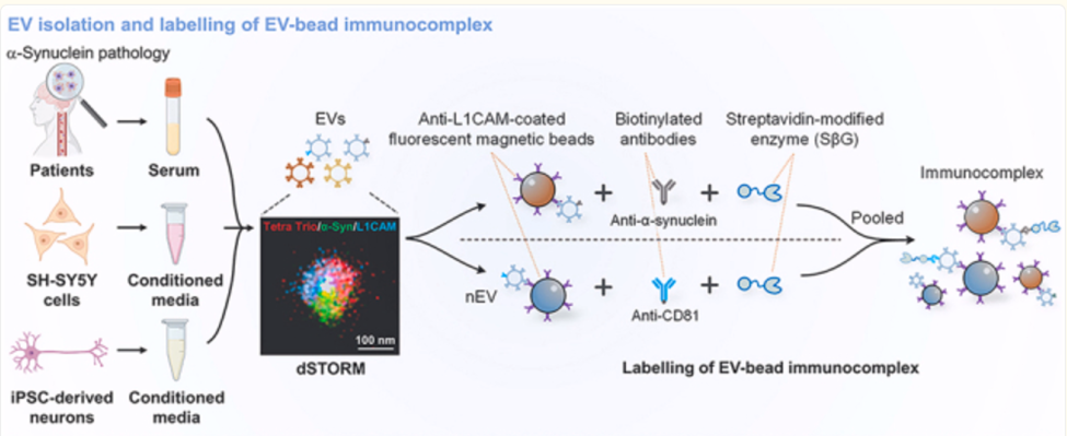

Representative image of a single EV obtained with dSTORM and corresponding quantification showing the % colocalization of L1CAM and α-Syn on Tetraspanin Trio+ (Tetra Trio+) EVs from SEC F3+F4; pooled serum from 5 individuals with PD was tested; scale bar, 100 nm. Reproduced from Cell Rep Med. 2025 Mar 7;6(3):101999. doi: 10.1016/j.xcrm.2025.101999
How ONI’s Nanoimager Makes This Possible
The ONI Nanoimager brings super-resolution microscopy (dSTORM, PALM) and single-particle analysis to the benchtop. In this Parkinson’s study, the system enabled researchers to:
- Detect and quantify individual EVs as small as 50–150 nm in diameter
- Simultaneously analyze multiple markers using multi-channel fluorescence imaging
- Assess co-localization of α-synuclein with neuronal and tetraspanin markers (e.g., CD63, L1CAM)
- Distinguish surface-bound vs. intravesicular signals via intact vs. permeabilized imaging workflows
This fine-grained characterization led to the discovery that membrane-bound α-synuclein on L1EVs was significantly elevated in individuals at high risk of developing PD, even before motor symptoms appear.
The Oxford study shows that the ability to visualize and quantify biomolecules at the single-vesicle level is not just a technical advancement, It’s a paradigm shift in early disease detection. Tools like the ONI Nanoimager are turning concepts like liquid biopsy for neurodegeneration into reality.
As the exclusive channel partner for ONI in Asia and China, DKSH Life Sciences plays a key role in bringing this powerful imaging platform to researchers. Our deep technical knowledge and regional footprint allow us to:
- Facilitate hands-on demonstrations and proof-of-concept studies for your specific samples
- Offer expert application training and support to ensure success from day one
- Work with clinical and academic groups to integrate super-resolution workflows into EV, biomarker, and neurodegeneration research
- Support collaborative projects through workshops, knowledge-sharing events, and publication guidance
By lowering the barrier to nanoscale imaging, we help researchers unlock insights that were previously out of reach and in doing so, accelerate the journey from discovery to clinical impact. Contact DKSH to learn more about ONI’s Nanoimager and how it can empower your next breakthrough.